Physician Assistant

A Rare Case of Gliomatosis Cerebri Uncovered After Vertigo: Diagnostic Challenges in Diffuse Gliomas

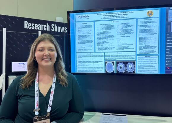
Photo of Caroline Butt standing in front of TV screen displaying poster project

Physician Assistant

A Rare Case of Gliomatosis Cerebri Uncovered After Vertigo: Diagnostic Challenges in Diffuse Gliomas

Caroline Butt ’26, MHS completed this project for PY 652: Medical Writing & Biostatistics for PA under the mentorship of Kara Strippoli MHS, PA-C, clinical assistant professor.

Overview

Gliomatosis cerebri is a rare and aggressive glial tumor characterized by diffuse infiltration of at least three cerebral lobes without forming a well-defined mass. Its widespread growth limits surgical options and reduces the effectiveness of standard therapies, highlighting the difficulty in diagnosis and management of this aggressive and uncommon form of diffuse glioma. This case demonstrates the unusual combination of widespread tumor infiltration and often minimal or absent neurological symptoms, making it a particularly challenging and complex neuro-oncologic scenario.

Rsearcher

Headshot of Caroline Butt

Caroline Butt ’24, MHS ’26

Physician Assistant, Master’s (Entry-Level Program)

School of Health Sciences

A Rare Case of Gliomatosis Cerebri Uncovered After Vertigo: Diagnostic Challenges in Diffuse Gliomas


Introduction

  • Gliomatosis cerebri is a rare and aggressive glial tumor characterized by diffuse infiltration of at least three cerebral lobes without forming a well-defined mass.1,2
  • The disease may present in two morphologic forms: a classical type characterized by diffuse infiltration without a discrete mass, and a secondary type exhibiting both diffuse involvement and a focal tumor component.6
  • The condition poses a diagnostic challenge due to its nonspecific presentation, which may include cognitive or behavioral changes, seizures, and progressive neurological deficits depending on the regions affected.7
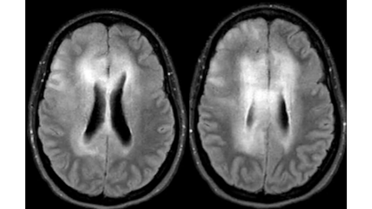
  • Imaging typically reveals widespread T2/FLAIR hyperintensity with minimal enhancement or mass effect, reflecting diffuse tumor infiltration rather than localized expansion.4
  • The diffuse pattern makes complete surgical resection nearly impossible, leaving radiotherapy and chemotherapy as primary treatments, often with limited effect.3,5,8
  • Prognosis is poor, with 26-52% surviving less than a year from symptom onset.7

Case Description

History of Presenting Illness

54-year-old female presented with 3 days of dizziness most consistent with vertigo and intermittent headache. The dizziness was worse with turning her head in certain directions and with laying down which caused her to feel like she was spinning in circles and nauseous.

Past Medical History 
  • PMH: Hashimoto's thyroiditis
  • PSH: Cesarean Section, hysterectomy with left oophorectomy due to fibroids (2015) and laparoscopic cholecystectomy
  • Allergies: NKDA
  • Medications: meloxicam 15 MG PO daily, metronidazole 0.75% vaginal gel 37.5 mg into the vagina nightly and synthroid 88 PO daily
  • Social History: 4 alcoholic beverages a week (2 glasses of wine, 2 cans of beer per week). No tobacco/illicit drug use.
Physical Exam
  • General: A&0x3, comfortable, no acute distress, speech clear and fluent, affect/mood normal
  • Heart: RRR, no murmurs/rubs/gallops
  • Lungs: Clear to auscultation bilaterally
  • Neuro: CN II-XII: PERRLA, EOM intact with some slight nystagmus on R end gaze, visual fields and acuity intact, facial sensation intact, face symmetrical with normal eye closure and smile, hearing normal to rubbing fingers, shoulder shrug intact
    • DH maneuver with L ear down provokes no symptoms or nystagmus. DH maneuver with R ear down provokes intense spinning vertigo and rotatory nystagmus
    • Coordination: FNF, HKS intact
    • Gait: tentative but stable and independent.
  • Vitals: BP 139/85 mmHg, HR 90 bpm, T 96.7 °F, RR 17 breaths/min, SpO2 98% on RA, BMI 29.48 kg/m^
  • Laboratory Analysis
    • WBC 9.4 x 10³/µL, Hgb 12.9 g/dL, Hct 39.3%, PLT 257 x 103/µL
    • TSH 2.41 mIU/L, ESR 22 mm/hr, RF <10 IU/mL, C-Reactive
Protein <0.3 mg/dL, ANA positive, SSA <1.0, SSB <1.0
    • CSF Analysis: Culture negative, Total Protein 33 mg/dL, Glucose 61 mg/dL
    • Meningitis/encephalitis PCR panel: Not detected
Differential Diagnosis
  • Neoplasm:
    • Glioma
    • Metastatic tumor
    • CNS lymphoma
  • Infection:
    • Brain abscess
    • Subacute viral encephalitis
    • Progressive Multifocal Leukoencephalopathy
    • Tuberculosis
  • Autoimmune/Inflammatory process
    • Tumefactive multiple sclerosis
    • Acute disseminated encephalomyelitis (ADEM)
  • Recent/acute infarct

Hospital Course

  • Physical examination revealed a normal physical and neuro exam and Epley maneuver was positive. Despite BPPV diagnosis, further testing was ordered as this was her first vertigo episode.
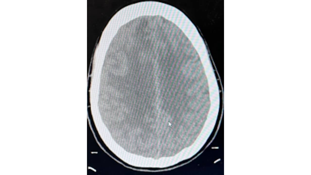
  • CT scan revealed a focal area of edema in the right frontal lobe suspicious for an neoplasm or abscess.
  • Pt was admitted and MRI revealed extensive T2/FLAIR hyperintensity in the right frontal lobe, peri-insular regions, corpus callosum, and cingulate gyri.
  • Due to MRI findings, further testing was performed. LP was performed to reveal no abnormalities within the CSF fluid. All infectious and inflammatory testing that was drawn resulted with normal findings.
  • Brain biopsy was performed and the suspicious area within the right frontal lobe was noted to have extremely friable tissue. Frozen section was sent to pathology showing proliferation of glial cells but with no definitive diagnosis.
  • Permanent specimens were sent for further cytology with later resulted as glioma, mild pleomorphism, no mitotic figures or necrosis, IDH wildtype.

Diagnostics

  • CT Head: Vasogenic edema right frontal lobe compatible with underlying neoplasm or abscess.
  • MRI w/wo contrast: Extensive T2 FLAIR signal hyperintensity within the right frontal lobe, bilateral peri-insular region (right worse than left), along the corpus callosum and cingulate gyri bilaterally.
  • Brain Biopsy: Area of lesion noted to have extremely friable tissue. Frozen section sent to pathology showing proliferation of glial cells. Permanent specimens later resulted as glioma, mild pleomorphism, no mitotic figures or necrosis, IDH wildtype.


Case Outcomes

  • Diagnosis was confirmed to be gliomatosis cerebri due to the pathology findings and the widespread nature found on MRI.
  • Patient was discharged with Oncology/Neuro-Oncology follow-up for treatment planning.

Discussion

  • According to the National Cancer Institute, gliomatosis cerebri is no longer considered a separate diagnosis, but rather a description of the aggressive, diffuse pattern of growth of a glioma.9
  • The rarity of Gliomatosis Cerebri makes it difficult to define prognostic factors and appropriate management.8
  • Gliomatosis cerebri is biologically related to glioblastoma multiforme. However, while glioblastoma multiforme typically presents in older adults, gliomatosis cerebri more often affects children and younger adults. Gliomatosis cerebri is also far more diffuse, which makes treatment challenging despite generally being less overtly malignant.9

Conclusion

  • Although gliomatosis cerebri is extremely rare, with fewer that 100 cases diagnosed annually, it should remain on the differential given its nonspecific presentation.
  • This case underscores the need for continued evaluation when the diagnosis is uncertain and the essential role of imaging and further testing in achieving an accurate diagnosis.


Faculty Mentor


For Further Discussion

This serves as an overview of the project and does not include the complete work. 

Course Overview

PY 652: Medical Writing & Biostatistics for PA introduces biostatistics, evidence-based medicine, as well as critical review and application of evidence to clinical decision-making. Students learn to construct clinical questions and perform literature searches. Methods for critically appraising the literature and strategies for maintaining currency of medical knowledge through journal clubs are practiced. Review of basic techniques of medical writing and presentation allow students to develop presentations, posters and journal articles while incorporating peer review feedback.

Explore Our Areas of Interest

We've sorted each of our undergraduate, graduate and doctoral programs into unique Areas of Interest. Explore these categories to discover which programs and delivery methods best align with your educational and career goals.

Explore Health and Medicine at Quinnipiac