
Colonial coffins provide radiologic sciences students window to the past
August 02, 2017
August 02, 2017
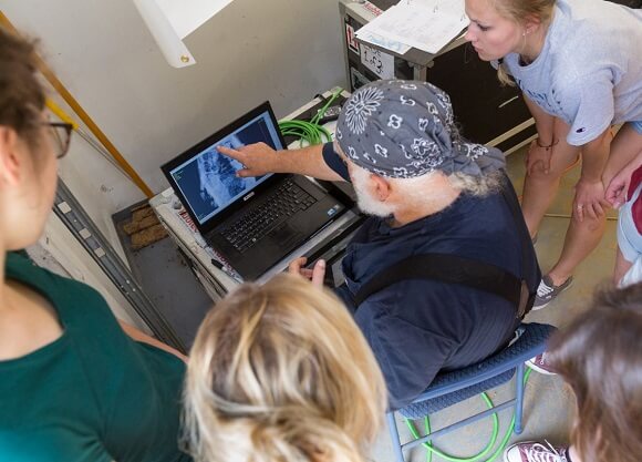
The Quinnipiac students traveled to Burlington, New Jersey with students from the University College Dublin, where they took X-rays of coffins and human remains dating as far back as the early 1700s. The coffins were a major archaeological discovery, and made national headlines when they were excavated from a construction site in the Old City neighborhood of Philadelphia in March.
“They are more than just boxes of bones,” said Gerald Conlogue, professor emeritus of diagnostic imaging and director of the Bioanthropology Research Institute at Quinnipiac. “They provide us with a picture of both a culture and a time period.”
Conlogue, who led the trip, has been using medical imaging techniques to study mummified and skeletal remains and artifacts for decades. He has worked on archeological sites from Lithuania to Peru, and regularly brings students along on projects.
“I want to broaden their perspectives, and show them that there are many other fields they can take their skills into,” he said.
Emily Paul ’18, a radiologic sciences major, recalls the excitement of arriving to find “artifacts everywhere,” a full skeleton being recovered by archeologists and state-of-the-art imaging equipment waiting for her.
Bioanthropology Research Institute
“I dug right in, radiographing skulls,” Paul said. “It isn't often that we get to play around with our modality like this.”
Paul and her team made a breakthrough while examining a child’s coffin. Focusing on dental development and the presence of “pins” meant to fasten a swaddle, they were able to determine age, cause of death and exactly when the deceased was buried.
“It was such a privilege to be a part of a discovery like this,” said Irish radiography student Rachel Whelan.
Whelan and her three classmates were able to participate in the project as part of a five-week exchange project between the School of Health Sciences and UCD.
“Using radiography anywhere other than a hospital was a new thing for us,” Whelan said. “It was really thought-provoking, examining the lives of people from so long ago. We learned so much.”
Word of the project’s success has already spread. Conlogue’s students are scheduled to present their findings before the Society for American Archaeology next year.
“They’ll have scholars and researchers with PhDs asking them how to do something,” Conlogue said. “Students need opportunities like that. It gives them a significant step up on everyone else.”
Paul looks forward to presenting her team’s findings, and educating people about the many uses of non-traditional radiology.
“I hope to continue immersing myself in this type of imaging,” she said. “Not many people have the privilege of experiencing their major in this setting.”

Quinnipiac Today is your source for what's happening throughout #BobcatNation. Sign up for our weekly email newsletter to be among the first to know about news, events and members of our Bobcat family who are making a positive difference in our world.
Sign Up Now