
‘The whispers of mummies,’ stories untold
October 28, 2022
October 28, 2022
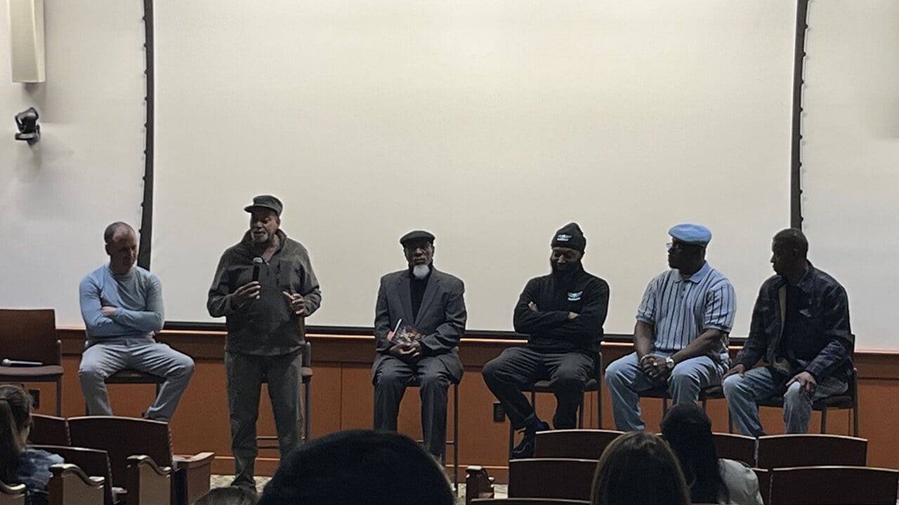
Geri Conlogue, professor emeritus, reflects on his experience traveling with fellow Professor Emeritus Ron Beckett, to Guanajuato, Mexico, to image a group of mummies in 2010.
“Most of us in this country have been, unfortunately, introduced to mummies via ‘horror’ movies,” said Conlogue. “Real mummies aren't frightening. Because of my clinical background, I see them as someone's relatives, a grandmother or grandfather. Also, my grandmother warned me to beware of the living and not the dead because the latter will not hurt you.”
Instead, Conlogue views mummies as passed individuals that offer insight on the history of the human species, learning that the concept of death is viewed differently all over the world.
“Mummies are actually the remains of people who walked the earth and experienced life during specific periods in history,” said Conlogue. “Locked within their bodies are hidden all the biological factors that affected their lives, for example, tuberculosis. Imaging, combining radiography and endoscopy, could be used to reveal those secrets to help provide an ‘eyewitness’ account of what life was like. Particularly with the poor segment of the population whose story may not have been written by a representative of that group.”
As a radiographer, Conlogue is trained in computed tomography (CT) and magnetic resonance (MR) imaging, both of which are required to provide diagnostic quality images for a radiologist. With that knowledge, Conlogue was able to modify his approach for looking at a live patient to one needed to produce diagnostic images of dehydrated mummified remains.
“For example, with a CT examine of a live patient, the goal is to use the least amount of radiation while sacrificing detail resolution,” said Conlogue. “However, with mummified remains, the resolution is the most important factor, disregarding radiation dose.”
Conlogue’s most recent experience imaging mummies in the same area of Mexico was six years ago.
“We partnered with a Stratford, Connecticut, manufacturer of x-ray equipment, Kubtec, to use their digital equipment to produce high-quality images of the remains,” said Conlogue. “Those images were recently used for an exhibit in the Mexican city.”
Conlogue tributes his further exploration of the field to his experience at Quinnipiac.
“When I came to Quinnipiac in 1992, I was given the ‘green light’ to include non-traditional imaging into the radiography curriculum,” said Conlogue. “With the encouragement of Ron Beckett, my chairman, one of the directions was imaging mummified and skeletal remains. If I was not at Quinnipiac none of this would have happened.”
Conlogue is currently working on another book with Ron Beckett titled “Side Show Mummies and Other Exhibited Remains” to educate people on this misconceived field of study.


Quinnipiac Today is your source for what's happening throughout #BobcatNation. Sign up for our weekly email newsletter to be among the first to know about news, events and members of our Bobcat family who are making a positive difference in our world.
Sign Up Now