
Pictures uncover the past
October 27, 2016
October 27, 2016
An MRI unit uses a body's natural magnetic properties to produce rich, detailed images. The process relies on hydrogen, which occurs in water and fat.
But what happens when you're trying to use the MRI on a mummy — which is dehydrated?
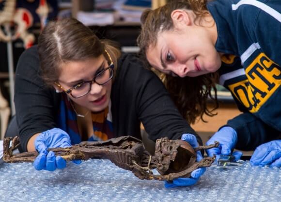
"You trick the MRI," said Emily Kelly, a diagnostic imaging student. During her senior year, that's what Kelly, fellow diagnostic imaging student Joy Pasqualucci ’16, and Bernadette Mele, clinical associate professor of diagnostic imaging, did to capture an image of a mummified infant, as well as a second mummy part — the arm of an adult woman.
A Seattle physician owned the mummies and, when he died, his wife donated them to the Mütter Museum of the College of Physicians of Philadelphia for an exhibit. The remains made a stop at Quinnipiac, where 30 students and several professors from different disciplines — including diagnostic imaging, radiologic sciences, anthropology and health science studies — used X-rays, MRI images, CT scans and good, old-fashioned examinations to learn more, such as age and cause of death.
Kelly and Pasqualucci placed a water bottle close to the mummy, and the MRI picked up the hydrogen in the bottle. With some calibrations, it also captured the magnetic materials of the infant's heart and veins.
"I gave them the challenge and they rose up and came up with a solution," Mele said.
The infant's heart may have been injected with a dye that had metallic properties, she said. In the 1800s, when the bodies were likely mummified, medical schools used them as teaching tools. They remain teaching tools for QU students.
Dental development tells age, for example, but without teeth, an MRI or CT scan is needed to see what's happening developmentally, said Jaime Ullinger, assistant professor of anthropology and co-director of the Bioanthropology Research Institute at Quinnipiac. "It's a great opportunity to put into action things they have done in class, and to see how you can apply what they have learned to different cases," she said. "It's also important that we have students from all different disciplines participating so they can see how we all work together."
Amel Langston, a health science studies student, said she was excited because she had never had this type of opportunity. She spent one class measuring the bones of the infant's body. "I was surprised how intact some of the features were, even after all this time," she said, pointing to the faint curve of the earlobe on the skull.
Larissa Mastroddi, a diagnostic imaging student who was part of the group that conducted CT scans, said she appreciated being able to examine mummified bodies, which isn't common at most schools. It showed another example of the different ways she could apply her skills beyond a traditional health care setting, she said.
Gerald Conlogue, professor emeritus of diagnostic imaging, said in a clinical setting, the diagnostic imaging equipment is programmed to work on human beings. In this situation, they have to look outside those boundaries to produce an image. "There isn't a button that says 'mummy.' This is a perfect real-world challenge for them. It gives them a better understanding of how this equipment operates."
"It's a great opportunity to put into action things they have done in class, and to see how you can apply what they have learned to different cases. It's also important that we have students from all different disciplines participating so they can see how we all work together." said Jaime Ullinger, assistant professor of anthropology and co-director of the Bioanthropology Research Institute


Quinnipiac Today is your source for what's happening throughout #BobcatNation. Sign up for our weekly email newsletter to be among the first to know about news, events and members of our Bobcat family who are making a positive difference in our world.
Sign Up Now