Quinnipiac University

Physician Assistant

Duodenal Hemorrhage and Hepatic Artery Occlusion Following Cholecystectomy

Graphic that says Duodenal Hemorrhage and Hepatic Artery Occlusion Following Cholecystectomy, Madison Villa, MHS ’26 Physician Assistant, Quinnipiac University, Hamden, CT

Physician Assistant

Duodenal Hemorrhage and Hepatic Artery Occlusion Following Cholecystectomy

Madison Villa, MHS ’26 completed this project as part of PY 652: Medical Writing & Biostatistics for PA under the mentorship of Sheree Piperidis, clinical professor of physician assistant studies.

Overview

This work by Madison Villa, MHS ’26 involves a case with a 48-year-old man that has a history of Mirizzi syndrome and a retained biliary stent from 2018 who presented to the ED with abdominal pain and leukocytosis. Through analysis of the patient's multidisciplinary management and recovery, the case highlights the importance of early recognition of atypical postoperative complications, collaborative care and long-term follow-up in complex biliary disease.

Researcher

Headshot of Madison Villa

Madison Villa ’24, MHS ’26

Physician Assistant, Master’s (Entry-Level Program)

School of Health Sciences

Duodenal Hemorrhage and Hepatic Artery Occlusion Following Cholecystectomy


Introduction

  • Subtotal cholecystectomy (SC) is indicated for complex gallbladder resections complicated by inflammation, infection, or distorted anatomy, obscuring the critical view of safety. 1-3
  • SC have minimal risk of post-operative complications; most common are bile duct injury or retained gallstones. 1-3
  • Biliary stents are effective for short-term management of stones or strictures, but lack long-term benefit due to risks of infection, bleeding, stent occlusion or migration, or duodenal perforation. 4
  • Evidence on retained biliary stents primarily comes from retrospective studies of patients lost to follow-up, who commonly presented with abdominal pain (94.7%), fever (52.6%), and jaundice (34.2%). 4
  • Actinomyces species are low-virulence anaerobes normally in the oral and genital mucosa that can cause granulomatous inflammation, abscess formation, and sinus tract development due to proteolytic enzymatic activity. 5,6
  • Hepatic actinomycosis is rare but may occur via hematogenous spread or prior surgery/foreign material; imaging often mimics malignancy but has a good prognosis and responds well to penicillin. 5,6
  • Post-operative hemorrhage after SC is uncommon, occurring in 0.3% of 1,231 cases in a meta-analysis from 1954-2013. 2
  • A meta-analysis of 2,918 laparoscopic SC from 1985-2020 demonstrated that incidence of vascular injury was only 0.1% and intestinal injury 0.2%. 3
  • Inadvertent duodenal injury during laparoscopic cholecystectomy is uncommon, but most likely to occur from cautery or dissection. 4
  • Bilio-enteric fistulas most often occur from chronic cholelithiasis, but rarely can be caused by intestinal ulcers, liver abscess, or iatrogenic damage. 8
  • Anatomical variations of the right hepatic artery occur in up to 25% of individuals and can create aberrancies near the triangle of Calot, increasing the risk of arterial laceration or thrombosis during biliary procedures. 9
  • Vascular complications from a laparoscopic cholecystectomy are extremely infrequent, but may arise from clip misplacement or thermal injury, leading to hemobilia or upper gastrointestinal bleeding. 10
  • When vascular injury occurs, the right hepatic artery is most often involved and increases rates of postoperative hemorrhage, hemobilia, and right-lobe ischemia. 11

Case Description

Case History:
  • 48-year-old man with PMHx of Mirizzi syndrome and a retained biliary stent from 2018 (lost to follow-up for removal)
  • No PSHx, allergic to penicillin (rash), no daily medications, remote h/o IVDU
  • Presented with LUQ abdominal pain and leukocytosis
  • US and CT with contrast showed cholelithiasis, intrahepatic and biliary dilation, and duodenal and pancreatic fat-stranding, suggesting diffuse inflammation
  • ERCP was done to remove the partial stent occlusion, sludge, and stones before a robotic subtotal fenestrating cholecystectomy
  • Intraoperatively, subhepatic and perigastric abscesses were drained, with cultures positive for Actinomyces and Streptococcus anginosus. Given the patient’s penicillin allergy, clindamycin was initiated.
  • Despite antibiotic treatment, WBC continued to rise, peaking at 32.4 cells/mm3 on POD6 preceding a massive 1L hematemesis with bloody RUQ drain output. 
Vitals:
  • BP: 111/76 mmHg
  • Pulse: 136 bpm
  • Temp: 99.1 °F (tympanic)
  • Respiration: 24 breaths/min
  • SpO2: 98% 5L NC
  • BMI: 31.81 kg/m2
DDX:
  • Persistent abscess/infectious erosion (due to ineffective antibiotic coverage)
  • Duodenal injury/cholecystoduodenal fistula
  • Surgical trauma/iatrogenic damage
  • Right hepatic artery pseudoaneurysm
  • Stress ulcer 
Physical Exam:
  • General: Alert and oriented, sitting in bed in mild discomfort, able to answer questions appropriately, pale and diaphoretic 
  • Pulm: Increased respiratory rate, no accessory muscle use or signs of respiratory distress. On 5L NC, 98%, CTA without crackles or wheezing
  • CV: Tachycardic to 130s, normal rhythm. No murmurs, rubs, or gallops
  • Abdomen: Actively vomiting large amounts of bright red blood and clots, diffusely TTP, no guarding or rebound tenderness, right abdominal Blake drain with bloody output
Diagnostics and Imaging:
  • WBC: 32.4 x 10³/µL (H)
  • Hb/HCT: 12.0 g/dL/36.5% (L)
  • Platelets: 845,000/μL (H)
  • Lipase: 101 U/L (H)
  • Alkaline phosphate: 179 U/L (H)
  • Remainder of CBC and CMP wnl
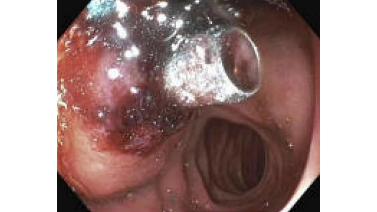
  • EGD: Anterior duodenal bulb bleed with an adherent clot and visible vessel. Friable and edematous tissue present suggestive of an impending cholecystoduodenal fistula
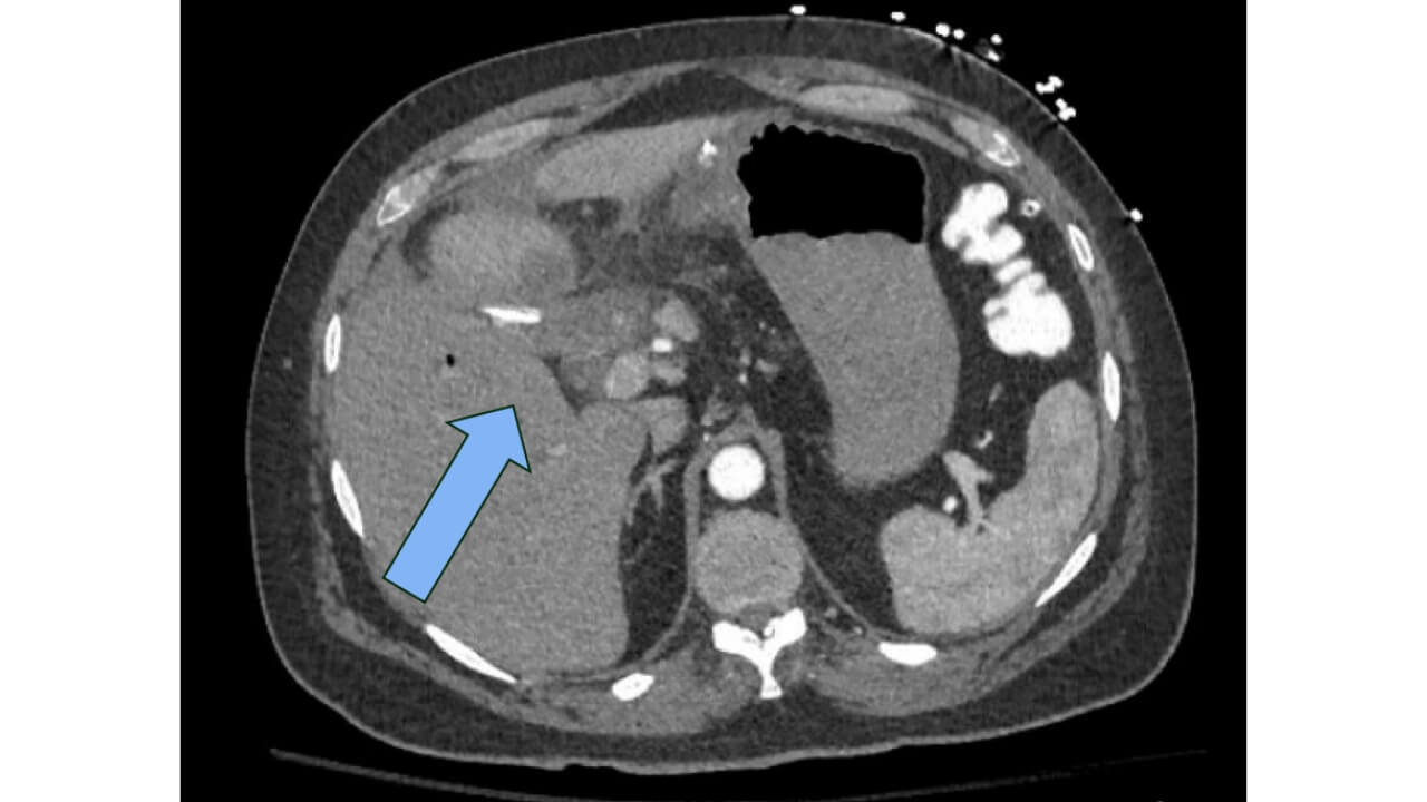
  • CTA Abdomen/Pelvis w/ Contrast: Filling defect of the right hepatic artery. No arterial extravasation. An almost complete occlusion of the right hepatic artery with minimal distal opacification
  • CT Abdomen/Pelvis w/ Contrast: Two hypoenhancing lesions within left hepatic lobe adjacent to gallbladder remnant measuring 1.6 x 1.4 cm and a small collection in the gastrohepatic region measuring 2.7 x 1.5 cm
Table 1: Post-operative trends of vital signs leading up to
hematemesis on POD6
POD Temperature (°F) WBC
(×10³/µL)
Hemoglobin
(g/dL)
Platelets
(×10³/µL)
1 101.2 17.8 12.5 543
2 100.9 18.5 12.1 567
3 100.3 19.8 12.1 579
4 100.2 19.9 12.1 629
5 99.2 22.9 11.9 631
6 98.9 32.4 12.0 845


Management

Emergent management of hematemesis:
  • MTP activation with 2 units of PRBC
  • IVF w/ LR @75 cc/hr
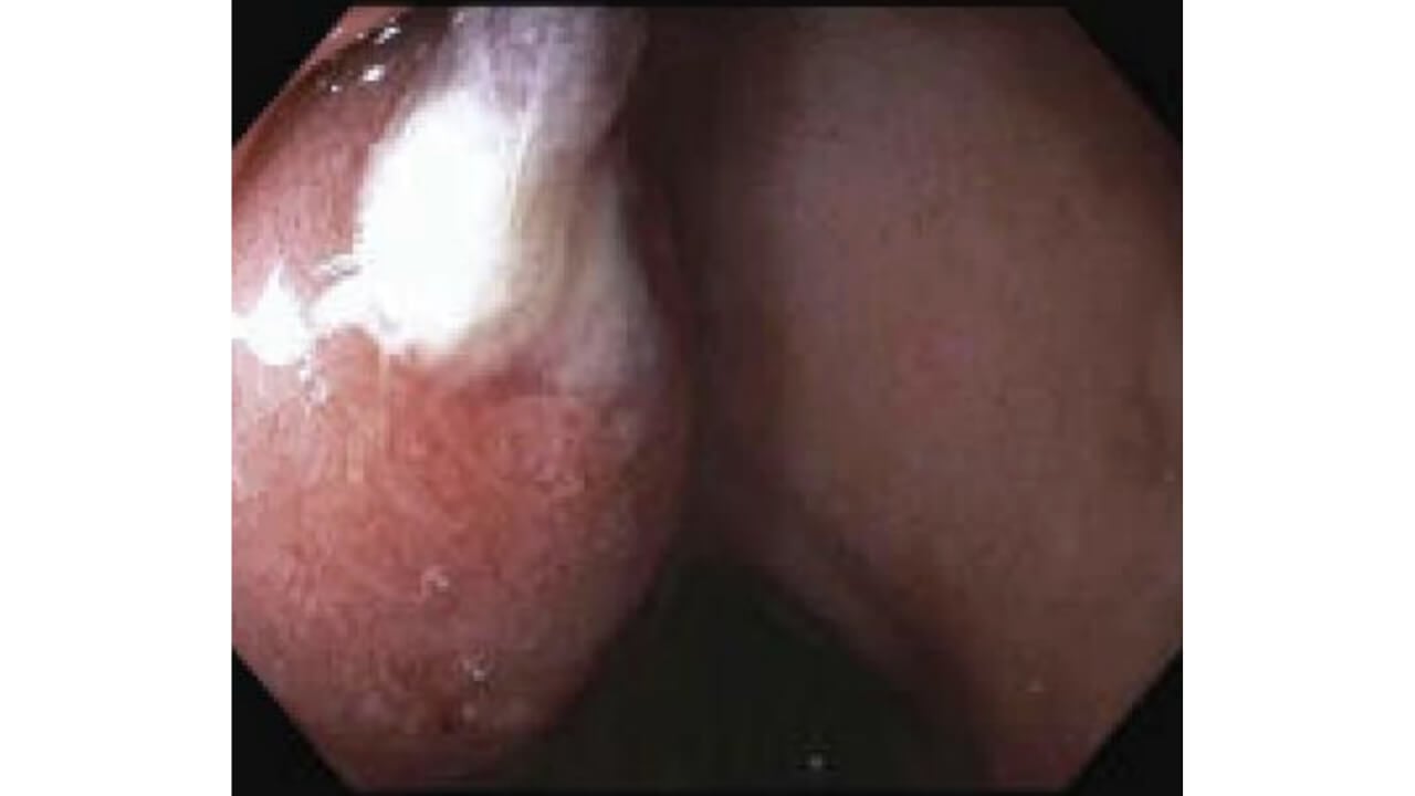
  • Emergent EGD with ligation of bleeding vessel using 3 hemoclips (two 16 mm Microtek clips, one 11mm Revolution 360) and NexPowder hemostatic agent
  • Transferred to SICU once stabilized 
Remainder of hospital course (7 days following  hematemesis, total hospital course of 20 days):
  • Returned to surgical floor one day following hematemesis 
  • Switched from clindamycin to IV ceftriaxone + metronidazole →PO cefpodoxime + metronidazole for 10 days upon discharge
  • NPO with NGT-LWS → clear diet → low fat diet upon discharge
  • Anticoagulation held until 3 consecutive stable H/H → IV heparin → apixaban PO BID on discharge
  • Discharged with outpatient vascular surgery follow up

Discussion

  • Retained biliary stents can promote ongoing inflammation, abscess formation, and mucosal erosion, providing a plausible pathway for Actinomyces infection, leading to indolent duodenal and biliary tissue damage. 4-7
  • The patient’s history of Mirizzi syndrome and recurrent cholelithiasis suggests longstanding biliary inflammation, increasing the risk of duodenal erosion and impending fistula development. 8,10
  • Although vascular complications of cholecystectomy are rare, the preexisting infection and difficult anatomy of this patient likely contributed to the postoperative hemorrhage and right hepatic artery occlusion. 1-3,9-11

Conclusion

  • Progressive leukocytosis, anemia, and thrombocytosis (Table 1) were the only indicators of the impending hemorrhage, emphasizing the importance of closely monitoring postoperative hemodynamics in complex biliary cases.
  • This case highlights the need for timely follow-up and having a heightened clinical awareness for serious complications when inflammatory, infectious, and iatrogenic risk factors converge. 


Faculty Mentor

References

Byskosh A, Stocker B, Liu DY-Z, et al. Subtotal fenestrating cholecystectomy: A safe and effective approach to the difficult gallbladder. J Surg Res. 2023;282:191-197. doi:10.1016/j.jss.2022.09.014

Elshaer M, Gravante G, Thomas K, Sorge R, Al-Hamali S, Ebdewi H. Subtotal cholecystectomy for “difficult gallbladders.” JAMA Surg. 2015;150(2):159. doi:10.1001/jamasurg.2014.1219

Nzenwa IC, Mesri M, Lunevicius R. Risks associated with subtotal cholecystectomy and the factors influencing them: a systematic review and meta-analysis of 85 studies published between 1985 and 2020. Surgery. 2021;170(4):1014-1023. doi:10.1016/j.surg.2021.03.036

Sohn SH, Park JH, Kim KH, Kim TN. Complications and management of forgotten long-term biliary stents. World J Gastroenterol. 2017;23(4):622-628. doi:10.3748/wjg.v23.i4.622

Matin MM, Novikov A, Sheth N, et al. Hepatic actinomycosis: a rare culprit in liver infections. Am J Gastroenterol. 2023;118(10S):S2422 S2423. doi:10.14309/01.ajg.0000964664.62683.40

Chegini Z, Didehdar M, Tabaeian SP, Khoshbayan A, Shariati A. A systematic review of case reports of hepatic actinomycosis. Orphanet J Rare Dis. 2021;16(1). doi:10.1186/s13023-021-01821-5

Machado NO. Duodenal injury post laparoscopic cholecystectomy: incidence, mechanism, management and outcome. World J Gastrointest Surg. 2016;8(4):335-344. doi:10.4240/wjgs.v8.i4.335

Stagnitti F, Tudisco A, Ceci F, et al. Biliodigestive fistulae and gallstone ileus: diagnostic and therapeutic considerations. Our experience. G Chir. 2014;35(9-10):235-238

Babbar V, Biswas R, Kumar A, et al. Right hepatic artery pseudoaneurysm following laparoscopic cholecystectomy: a rare complication treated with coil embolization. Cureus. Published online July 2025. doi7759/cureus.89125

Ullah K, Baig MA, Jan Z, Nazar MW, Shardha HK, Kumar G. Post-laparoscopic cholecystectomy extra-hepatic arterial pseudoaneurysm: an uncommon complication. Int J Surg Case Rep. 2022;95. doi:10.1016/j.ijscr.2022.107221

Stewart L, Robinson TN, Lee CM, Liu K, Whang K, Way LW. Right hepatic artery injury associated with laparoscopic bile duct injury: incidence, mechanism, and consequences. J Gastrointest Surg. 2004;8(5):523-531. doi:10.1016/j.gassur.2004.02.010


Further Resources

This serves as an overview of the project and does not include the complete work. 

Course Overview

PY 652: Medical Writing & Biostatistics for PA introduces biostatistics, evidence-based medicine, as well as critical review and application of evidence to clinical decision-making. Students learn to construct clinical questions and perform literature searches. Methods for critically appraising the literature and strategies for maintaining currency of medical knowledge through journal clubs are practiced. Review of basic techniques of medical writing and presentation allow students to develop presentations, posters and journal articles while incorporating peer review feedback.

Explore Our Areas of Interest

We've sorted each of our undergraduate, graduate and doctoral programs into unique Areas of Interest. Explore these categories to discover which programs and delivery methods best align with your educational and career goals.

Explore Health and Medicine at Quinnipiac